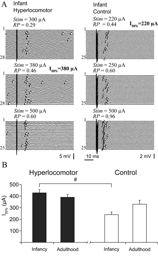
Figure 4. Impaired striatal responsiveness to cortical electrical stimulation in infant hyperlocomotor mice.
A. Typical responses of striatal single units to cortical stimulation in infant hyperlocomotor (left) and control (right) mice, at increasing stimulation currents (Stim). Response probability (RP) at different stimulation currents was estimated on the basis of 20 to 30 stimulation trials, as illustrated. B. The current needed to drive spike discharges in approximately 50% of the trials (I50%) was different between infant hyperlocomotor and control mice (significant interaction in a two way ANOVA, F(1,84)=5.71, p=0.019; # Tukey post-hoc test p<0.0003), but not in adulthood. Note that, because the relationship between stimulation current and probability of driving spikes did not seem linear, and only a limited number of current intensities could be tested for each neuron, the current we call I50% is in fact the tested current that drove spikes in approximately 50% of the trials. The average response probability corresponding to the intensities computed for the I50% parameter was as follows (mean ± SEM): 6-OHDA lesioned infant hyperlocomotor: 0.51 ± 0.04, infant controls: 0.55 ± 0.04, 6-OHDA lesioned adults: 0.56 ± 0.04, adult controls: 0.58 ± 0.06. Response probability at I50% was not different between groups (two way ANOVA). For neurons that were responsive to both cortical areas, only the minor I50% was used to study striatal sensitivity to cortical stimulation. These data correspond to mice of set one.

