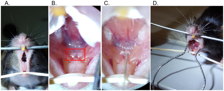
Figure 2. Retrograde injection steps.
A. Access the oral cavity by separating the maxillary and mandibular incisors. B. Visualize the papillae (boxed) below the tongue at the floor of the mouth, which mark the location of Wharton’s duct. C. Using a catheter with wire inset, gently cannulate the base of the submandibular papilla. D. Following cannulation, the catheter tubing can be exchanged with syringe tubing

