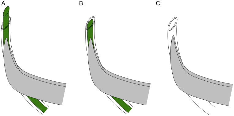
Figure 3. Effective positioning of catheter and stylet for Wharton’s duct cannulation.
A. Align the tubing with the curvature of the forceps, and cut a beveled end on the tubing and wire to initially puncture the sublingual papilla. B. Retract the stylet within the tubing to make a rigid guide to insert the tubing within the sublingual papilla. C. Insert catheter tubing (stylet removed), joined to injection syringe, within the previously made orifice.

