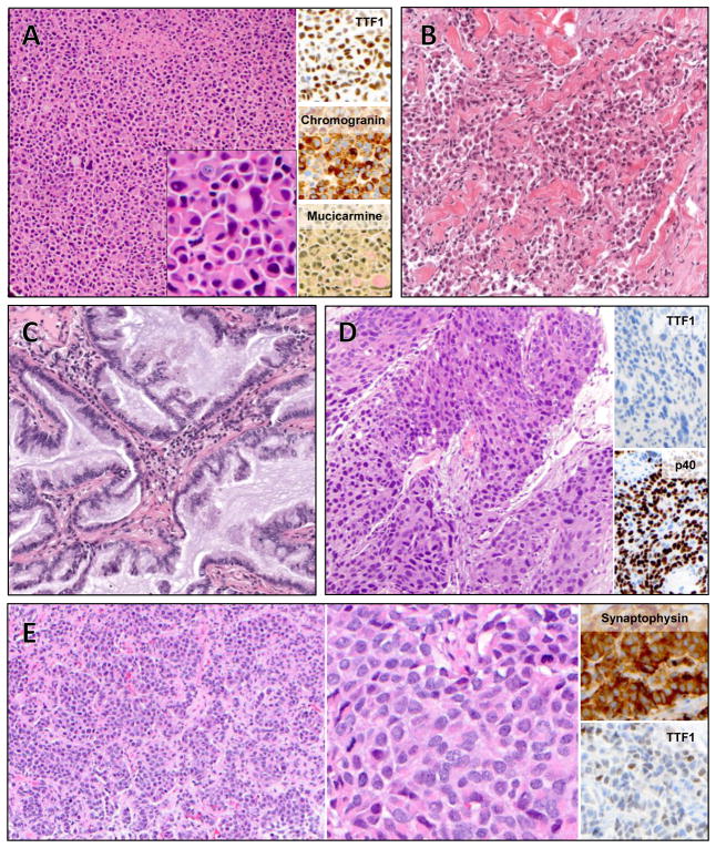
Figure 2.
Histology of select cases. A: Case 2, adenocarcinoma with solid growth pattern, diffuse neuroendocrine differentiation, and signet ring cells. Inset, high magnification. B: Case 1, poorly differentiated adenocarcinoma, with solid and single-cell growth patterns. C: Case 4, mucinous adenocarcinoma. Case 6 had similar histology (not shown). D: Case 8, squamous cell carcinoma. E: Case 11, neuroendocrine carcinoma with well-differentiated morphology and increased mitotic activity (left) and high power (middle).

