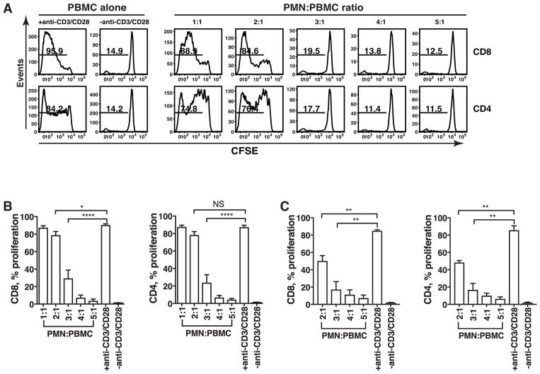
Figure 1. PMN inhibit T cell proliferation in a dose-dependent manner.
CFSE-labeled PBMC stimulated with or without anti-CD3/CD28 mAbs were co-cultured with increasing numbers of fixed PMN (Fig 1A, 1B) or unfixed PMN (Fig. 1C) for 5 days. Flow cytometry was used to determine CFSE dilution in live CD8 and CD4 T cells. (A) The result is a representative of nine different donors. Numbers on histograms represent the percentage of proliferating T cells. (B) Results from nine different donors (n =9) are expressed as mean ± SEM. ****P < 0.0001, *P < 0.05, P =NS (not significant). (C) CFSE-labeled PBMC were co-cultured with unfixed PMN in the presence of anti-CD3/CD28 mAbs at indicated ratios. Results are shown as mean ± SEM (n =3) from three different donors. **P < 0.01.

