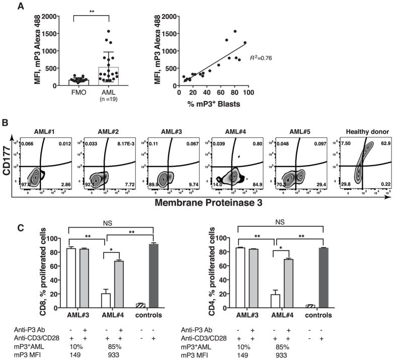
Figure 7. mP3+ AML mediates the inhibition of T cell proliferation, and proliferation is restored via P3 blockade.
(A) Flow cytometric analysis of CD177 and mP3 expression on AML blasts and healthy donor PMN were performed. FMO, fluorescence minus one. (B) Representative plots of AML blasts from AML patients (n =19) and PMN of healthy donor (n =20) are shown. (C) Following fixation with 0.5% PFA, allogeneic AML were pre-incubated with or without anti-P3 Ab for 30 minutes before they were co-cultured with PBMC at a 5:1 ratio (experiment was performed in duplicates) for up to 5 days. PBMC stimulated with or without anti-CD3/CD28 mAbs were used as positive and negative controls, respectively. Error bars represent mean ± SEM. **P < 0.01, *P < 0.05, P =NS (not significant).

