
Figure 2. Generation of primary recipient mice by lentiviral transduction of transgenic TRF2 into GFP and CD45.1 donor bone marrow.
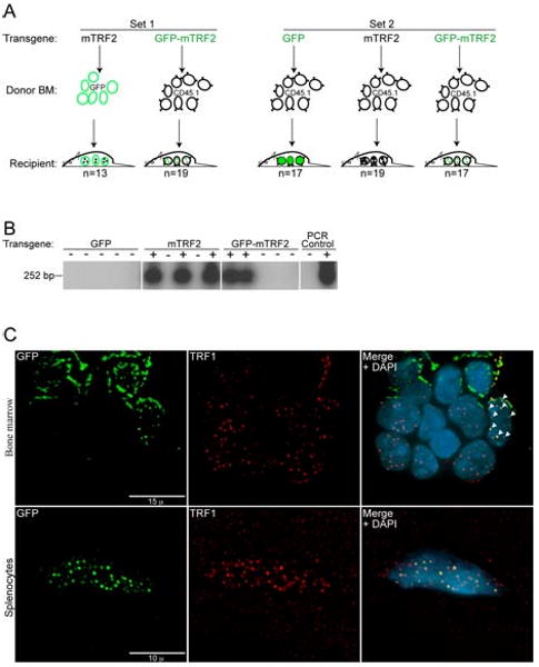
(A) A primary #1 mouse colony (Set 1) was generated by the transduction of GFP donor bone marrow (BM) with the mTRF2 transgene and CD45.1 donor bone marrow with the GFP-mTRF2 transgene. For the primary #2 colony (Set 2) only CD45.1 donor bone marrow was used and transduced with a GFP-control, the mTRF2 or the GFP-mTRF2 transgene. Recipient mice were C57BL/6J. (B) Genotyping of primary recipient mice transduced with transgenic GFP-mTRF2 and GFP by nested PCR and subsequent Southern analysis. Examples for Set 1 and Set 2 are displayed. As a negative control (-) genomic DNA from a C57BL/6J mouse was used. As a positive PCR control (+) genomic DNA of GFP-mTRF2 expressing HeLa cells was included. (C) Transgenic GFP-mTRF2 is expressed in the hematopoietic system of recipient C57BL/6J mice. Indirect immunofluorescence of bone marrow (top) and spleen (bottom) isolated from a secondary recipient of GFP-mTRF2 expressing bone marrow. GFP-mTRF2 was visualized by the GFP-tag, TRF1 was detected by a mTRF1 specific antibody. Chromatin was counterstained with DAPI. White arrows indicate co-localization of endogenous TRF1 with recombinant GFP-mTRF2.