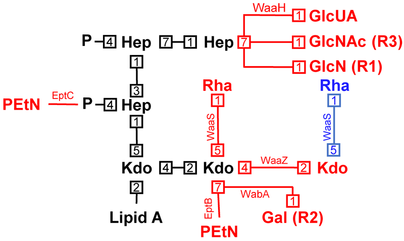
Fig. 7: Modifications to the structure of the core oligosaccharide.
Shown is the conserved inner core oligosaccharide (and its linkage to lipid A) in black, with potential modifications being indicated in red (though the alternate rhamnose modification by WaaS when the second Kdo is modified with PEtN by EptB is shown in blue). Numbers represent bond positions between sugars. Enzymes mediating modifications are next to each linkage, and when not associated with E. coli K-12, core type associations are listed in parentheses beside the modification.

