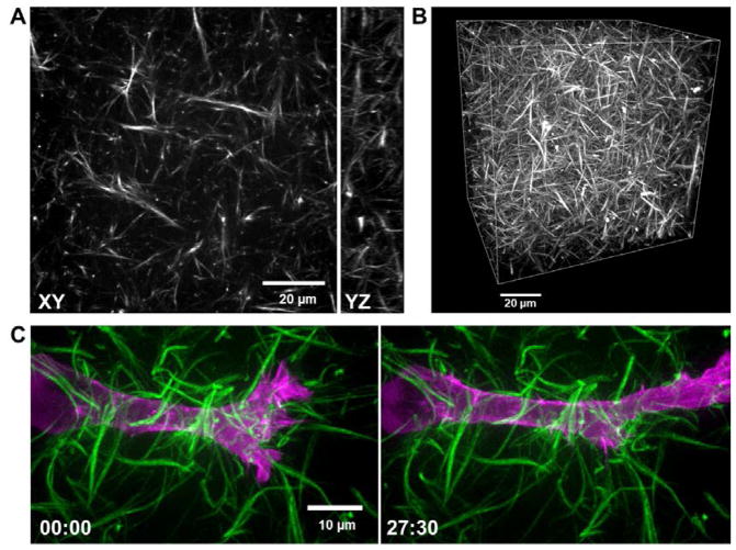
Figure 2. Fluorescence images of a 4% labeled collagen.
A. Single confocal slice (XY) and a 10 μm YZ projection (right) of a 4% atto 488-labeled 3 mg/ml rat-tail collagen gel polymerized at 21 °C. B. 3D rendering of the collagen gel shown in panel A (X, Y, Z dimensions: 100 × 100 × 80 μm, respectively). C. High-resolution max intensity projections of an HT-1080 fibrosarcoma cancer cell transfected with EGFP-α-actinin (magenta) migrating through Atto 565-labeled 3 mg/ml rat-tail collagen (green) polymerized at 16 °C. Images show first frame (left) and 63rd frame (right) in a time-series (imaged every 0.5 μm in Z over 15 microns, every 30 sec). Time is in minutes.

