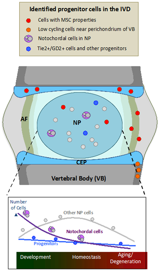
Figure 2:

Identified progenitor cells populating the IVD and the changes in their numbers as a function of time, from development, through homeostasis and to aging. Depiction of cells with MSC properties, low cycling cells near the perichordium of the vertebral body, notochordal and Tie2+/GD2+ and other progenitor cells.
