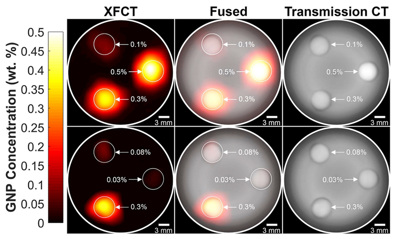
Figure 3.

Reconstructed and smoothed XFCT (left column), transmission CT (right column), and fused (middle column) images of 3-cm-diameter PMMA phantom under both the “high” (top row) and “low” (bottom row) configurations. The concentrations of the GNP/PBS inserts for the “high” configuration were 0.1, 0.3, and 0.5 wt. % and 0.03, 0.08, and 0.3 wt. % for the “low” configuration.
