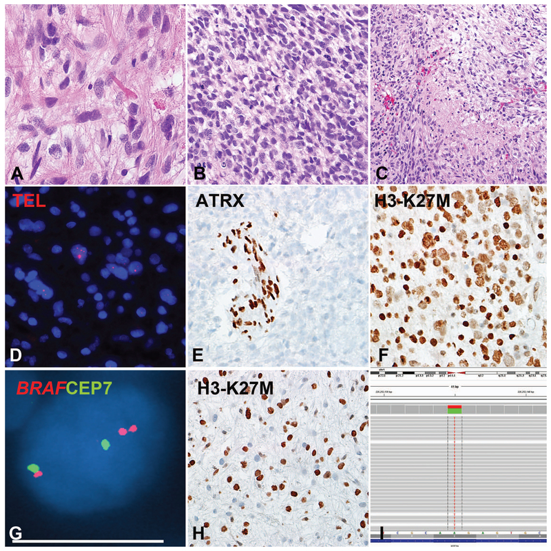
Figure 3: Pilocytic astrocytoma with anaplasia (PA-A) with concurrent BRAF duplication, ALT and H3-K27M mutation (Case 9).
This PA-A developed in the cerebellum of a 53-year-old woman during progression from a pre-existing PA. The precursor and the most recent resection had compact piloid areas with Rosenthal fibers and eosinophilic granular bodies (A). The anaplastic component had brisk mitotic activity (B) and pseudopallisading necrosis (C). Molecular alterations detected included ultrabright telomere FISH signals indicating ALT (D), ATRX loss (E), expression of H3-K27M mutant protein in almost 100% of neoplastic cells (F) and BRAF duplication (G). H3-K27M expression (H) and ATRX loss (not shown) were also present in a precursor 2 years prior, although the latter appeared to be present at a lower frequency in neoplastic cells. Next generation sequencing detected a AAG->ATG missense mutation in H3F3A at codon 27 Lysine (K) that was changed to Methionine (M) in all three H3-K27M immunohistochemistry positive cases tested (I).

