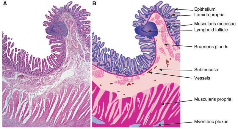
Figure 1.
Small intestinal mucosal architecture. (A) Low magnification image of hematoxylin and eosin-stained section of normal human duodenum. Many of the structural features that are common throughout the gastrointestinal tract and other mucosal surfaces can be appreciated. (B) Line diagram indicating specific structures that comprise the intestinal wall. The open spaces between fibers of the muscularis propria represent artefactual separation that occurred during tissue processing. (From Podolsky et al. 2015; reprinted, with permission from John Wiley & Sons ©2015.)

