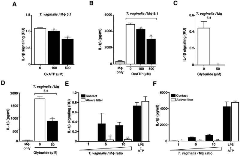
Fig. 3: Response to extracellular ATP via P2X7 receptor signalling and physical contact contributes to T. vaginalis inflammasome activation.

THP-1 macrophages were exposed to T. vaginalis at an MOI of 5 (T. vaginalis: host) for 4 h in the presence of (A-B) oxidized ATP (OxATP) or (C-D) Glyburide, an ATP-gated P2X7 receptor inhibitor. (E-F) A 0.4 μm membrane filter that allowed soluble materials to flow through but prevented contact was placed in between THP-1 macrophages and T. vaginalis in the above filter condition, or parasites were placed below the filter to allow contact between T. vaginalis and macrophages (contact). THP-1 cells were treated with 10 ng/ml LPS+ 5 mM ATP as a positive control for NLRP3 activation. Levels of bioactive IL-1β were detected using the IL-1β reporter cells (A, C, and E) and total IL-1β protein levels were detected by ELISA (B, D, and F). A representative result from 3 independent experiments is shown in A-D, and two independent experiments in E and F. Graphs show the mean with standard deviations. (A-D) **p-value<0.01 compared to THP-1 cells exposed to T. vaginalis in the presence of vehicle control. (E-F) *p-value<0.05, **p-value<0.01 compared to contact with T. vaginalis condition at that MOI.
