
Fig. 4: Inflammasome activation by T. vaginalis contributes to pyroptosis.
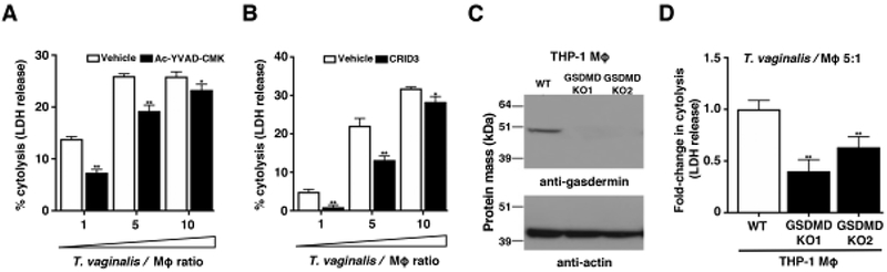
Cytolysis of host cells was assessed by measuring lactate dehydrogenase release from dying macrophages. (A-B) THP-1 macrophages were incubated with T. vaginalis using different multiplicities of infection (T. vaginalis: host cell) for 4 h in the presence of vehicle or (A) the caspase-1 peptide inhibitor Ac-YVAD-CMK or (B) the NLRP3/AIM2 inhibitor CRID3. Data are representative from 3 experiments. Graphs show the mean with standard deviations, *p-value<0.05, **p-value<0.01 compared to vehicle treatment at that multiplicity of infection. (C-D) Gasdermin D (GSDMD) was knocked out in THP-1 cells using CRISPR-Cas9. (C) Confirmation of gasdermin D knockout in two clones by western blot analysis using an anti-gasdermin D antibody. Actin loading control and molecular weight markers in kDa are also shown. (D) GSDMD KO cells or wildtype THP-1 cells were exposed to T. vaginalis at an MOI of 5 for 4 h. The average fold change in percent cytolysis compared to THP-1 wildtype cells from three independent experiments with standard errors of the mean are shown, **p-value<0.01 compared to THP-1 wildtype cells.