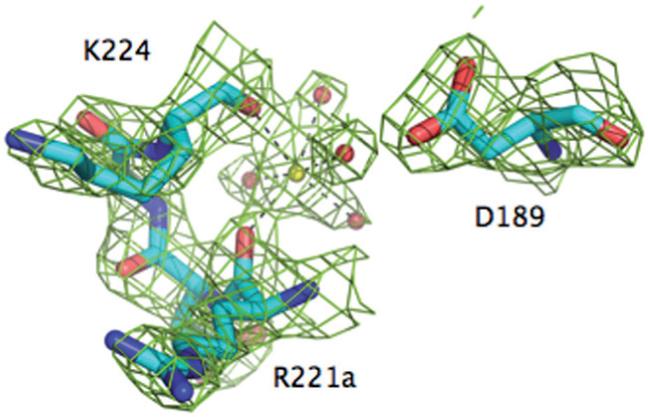
Figure 1.

The Na+ binding site of meizoIIRRΔF1. Shown is the 221a-224 segment of the 220-loop, along with the side chain of D189. Na+ (yellow ball) is coordinated octahedrally by the backbone O atoms of R221a (2.4 Å) and K224 (2.5 Å) and four buried water molecules (red balls, 2.5-2.6 Å). One of these water molecules connects to the Oδ2 atom of D189. The Na+ coordination shell of meizoIIRRΔF1 is practically identical to that of thrombin [20], underscoring the similarity of Na+ affinity and kinetic activation between the two enzymes (Table 1). The electron density 2F0-Fc map (green mesh) is contoured at 2 σ.
