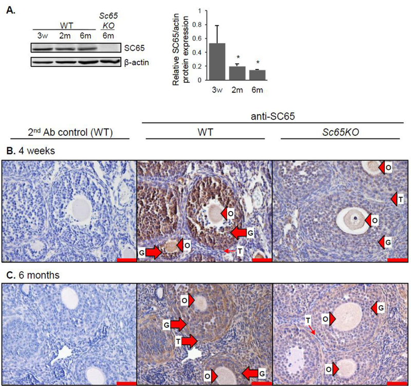
Figure 4.

SC65 is expressed in follicular granulosa cells at 4 weeks and in granulosa, theca and stromal cells in 6 month-old mouse ovary. A. Western blot analysis for SC65 in WT and Sc65KO mouse ovaries at the ages indicated. Quantification by densitometry is shown to the right, normalized to beta-actin, n = 3 replicates, * p < 0.05 compared to 3 weeks old. B. and C. IHC for SC65 on ovary from WT and Sc65KO mice at 4 weeks old (B) and 6 months old (C). Large arrows indicate positive cells, small arrows indicate negative cells, and arrowheads indicate non-specific staining. O: oocyte, G: granulosa cells, T: theca cells. Scale bars are 50 μm.
