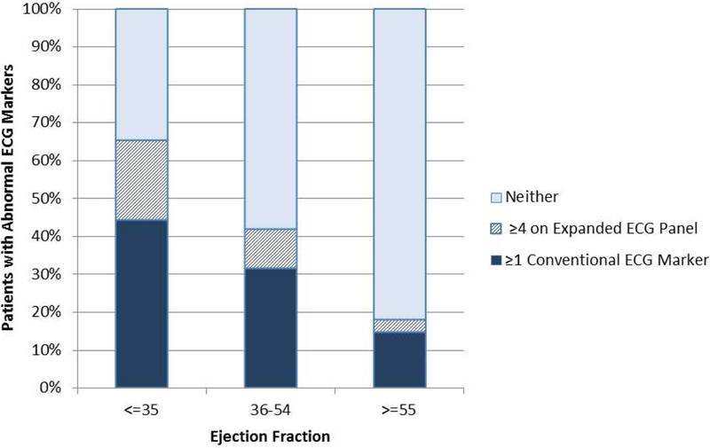
Figure 1. Association of abnormal ECG markers with LVEF.

Among all patients (n=9742), ≥1 conventional abnormal ECG marker was present in 44% of patients with LVEF ≤35% (in dark). Among patients without conventional ECG abnormalities, an additional 21% of patients with LVEF ≤35% had ≥4 abnormal markers on the expanded ECG panel (in cross-hatch). Use of the expanded panel plus conventional markers increased identification of abnormal ECG findings from 44% to 65% among patients with LVEF ≤35%.
Conventional ECG abnormalities were left bundle branch, atrial fibrillation/flutter, or paced rhythms.
The expanded abnormal ECG panel included resting heart rate >85 bpm, QRS duration >110 ms, QTc interval ≥460 ms for men and ≥470 ms for women, delayed QRS transition zone, delayed intrinsicoid deflection and QRS-T angle >90o.
