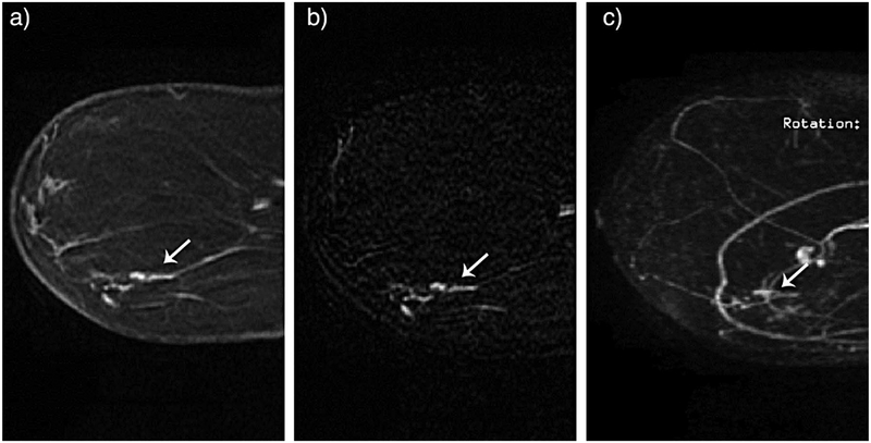
Figure 2.
54-year-old female for high risk screening. New linear nonmass enhancement in the lower outer left breast measuring 1.9 cm. MRI guided core biopsy yielded DCIS with microinvasion. Cancer recognized by all 4 radiologists on reading of the abbreviated protocol. First post-contrast sequence (A), first post-contrast subtraction sequence (B) and subtraction maximum intensity projections (MIP) sequence (C). Reprinted with permission from: Mango VL, Morris EA, Dershaw D, et al. Abbreviated protocol for breast MRI: are multiple sequences needed for cancer detection? Eur J Radiol. 2015;84(1):65–70.

