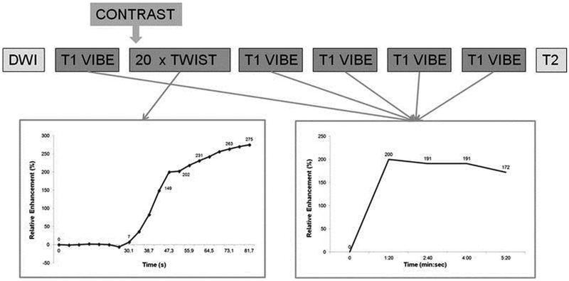
Figure 7.
Schematic drawing of the breast MRI scan protocol: The TWIST acquisitions allow evaluation of the contrast inflow in the lesion, whereas the VIBE acquisitions are used for 3 time point analysis, creating the classic contrast enhancement versus time curve. Reprinted with permission from: Mann RM, Mus RD, van Zelst J, et al. A novel approach to contrast-enhanced breast magnetic resonance imaging for screening: high-resolution ultrafast dynamic imaging. Invest Radiol. 2014;49(9):579–85.

