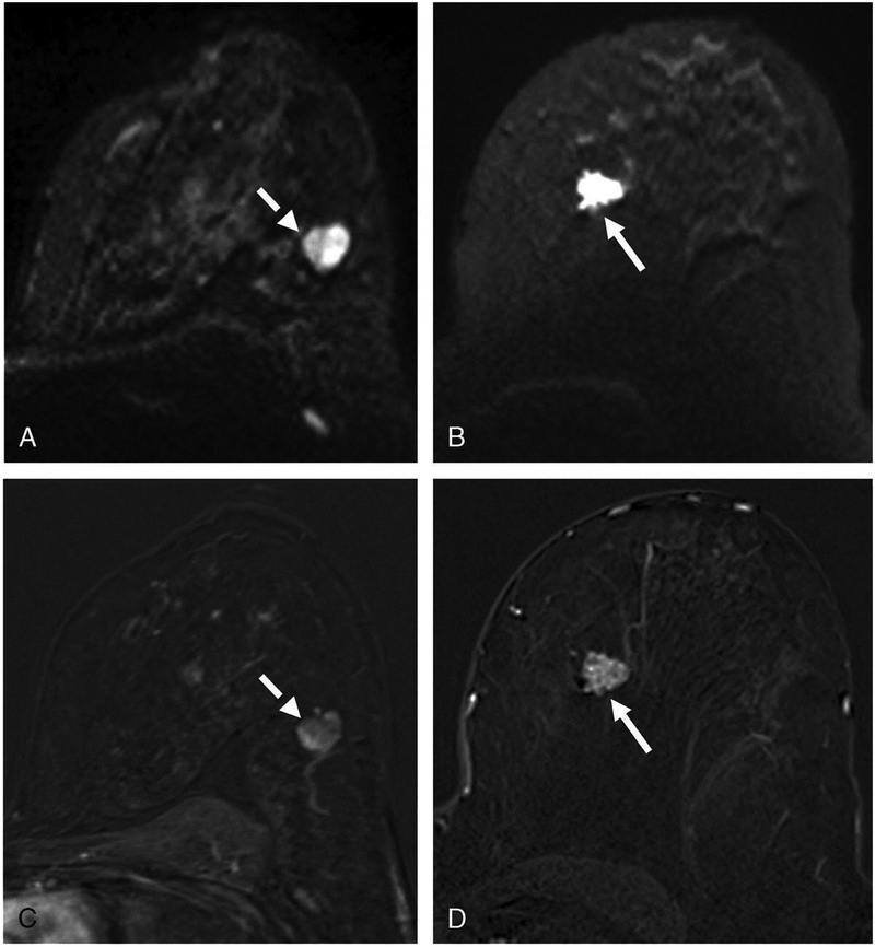
Figure 8.
Comparison of morphologic assessment on NC-MRI and CE-MRI. Panel A shows a circumscribed round lesion on the b850 s/mm2 image (dashed arrow) while panel B visualizes a noncircumscribed, rather spiculated lesion on the b850 s/mm2 image (arrow). The lesion from panel A appears with circumscribed margins on the early contrast-enhanced subtraction (C, dashed arrow) and was histologically proven as a fibroadenoma while the lesion from panel B appears noncircumscribed with heterogeneous internal structure and a feeding vessel on the early contrast-enhanced subtraction (D, arrow) and corresponds to an invasive ductal carcinoma G2. Reprinted with permission from: Baltzer PAT, Bickel H, Spick C, et al. Potential of Noncontrast Magnetic Resonance Imaging with Diffusion-Weighted Imaging in Characterization of Breast Lesions: Intraindividual Comparison With Dynamic Contrast-Enhanced Magnetic Resonance Imaging. Invest Radiol. 2018;53(4):229–235.

