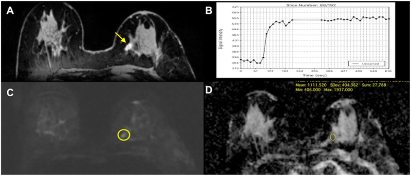
Figure 9.
Invasive ductal carcinoma (IDC) G1 in the left breast medial in a 71-year-old woman. (A) On DCE-MRI there is a 10mm irregular-shaped and marginated lesion (arrow) with (B) an initial fast/plateau enhancement (II); DCE-MRI findings were classified as suspicious for malignancy (BI-RADS 4). DWI was false negative as none of the readers called this lesion on DWI alone. However, when read as mpMRI combining DCE-MRI and DWI, readers identified a (C) hyperintense correlate (circle) with (D) ADC values measuring 1.111×10⁻³ mm2/s, which further confirmed malignancy.

