Fig. 4.
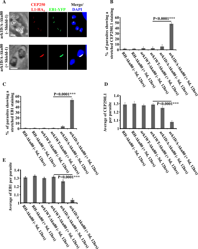
Interfering with TgArk1 function decreasingly affects the duplication of spindle pole and centrosome inner core. a Co-staining of TgCEP250L1-HA3 (a marker of the centrosome inner core, red dots) and TgEB1-YFP (spindle pole, green dots) shows under-duplication of both subcellular structures in the ark1D/A-Δku80 strain treated with shield-1 (lower panel). The ark1D/A-Δku80 strain in the absence of shield-1 (top panel) shows a normal pairing of spindle pole/centrosome inner core per nucleus (two spindle pole clusters/ two centrosome inner cores). The nuclei are stained with DAPI (in blue). Scale bars represent 2 μm. b, c Quantification of the number of parasites showing a stretched centrosome inner core or spindle pole in three different strains (RH-Δku80, ark1WT-Aku80 and ark1D/A- Aku80) in the presence or absence of shield-1 for 12 h. At least 900 parasites were examined for each condition. Values are mean ± SD for three independent experiments. d, e The average of TgCEP250L1- HA3 containing inner cores or TgEB1-YFP containing spindle poles per parasite, was quantified in 900 parasites revealing a significant reduction of the spindle poles and centrosomes inner cores when ark1D/A-Δku80 parasites were treated in presence of shield-1 (red and green dots). Values are mean ± SD for three independent experiments

