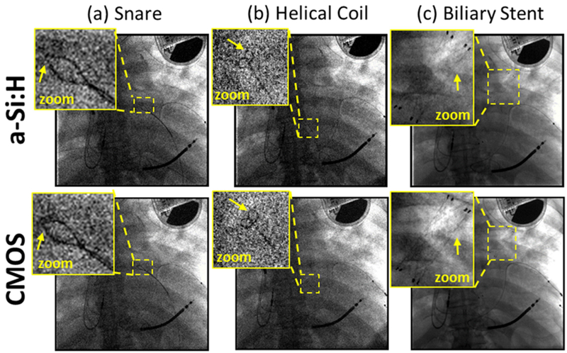
Figure 8.

Projection images of a cadaver and interventional devices. Images in (a) and (b) show a single frame (100 kV, 0.015 mAs, EAK ~11 nGy) from a fluoroscopic series during deployment of (a) a snare and (b) a helical coil. (c) Radiographic visualization of a stent (100 kV, 0.5 mAs equivalent, EAK ~0.4 μGy).
