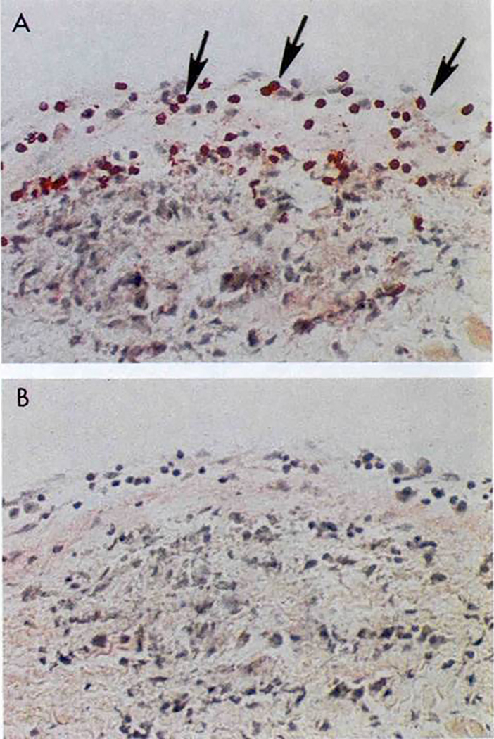
Figure 12.

Extravasation of CD4+ lymphocytes into valve above Aschoff’s body in the subendocardium of the left atrial appendage. Original magnification 200X Taken from Roberts et al (48). Section A stained with anti-CD4 Mab and Section B antibody isotype control.
