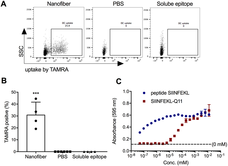
Figure 2. Peptide nanofibers displaying CD8+ epitopes are taken up by DCs in draining lymph nodes, and attached epitopes are presented within MHC-I.
Twenty hours after intranasal administration, TAMRA-labeled peptides in nanofibers were taken up by DCs in the draining mediastinal lymph nodes, whereas soluble peptides were not (A and B; Dose: 50μL of 2mM peptide). In vitro, SIINFEKL delivered as a soluble peptide and as SIINFEKLQ11 nanofibers was cross-presented by BMDCs in a dose-dependent manner, as detected by B3Z CD8+ hybridoma T cells that specifically recognize SIINFEKL presented in MHC-I H-2Kb (C). Data shown were combined from two independent experiments, with n=6 for PBS and n=4 for nanofibers and soluble epitopes in A and B. One representative experiment of 3 repeats is shown in C. Data are presented as mean±SD. Statistical significance was tested by one-way ANOVA and Tukey’s multiple comparison (*** p<0.001).

