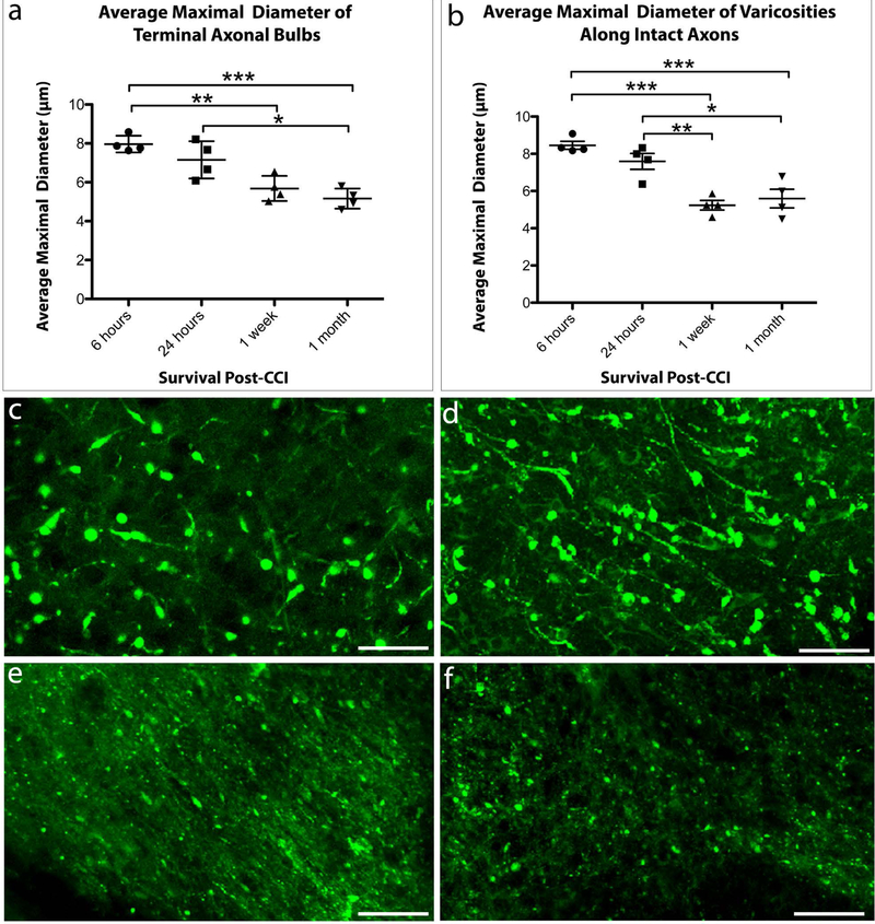
Figure 4. The Size of Axonal Swellings Decreases with Increased Survival Post-CCI.
The average maximal diameter of axonal swellings of both (a) terminally disconnected bulbs and (b) axonal varicosities along intact axons decreases over time following CCI. (*p ≤ 0.05, **p ≤ 0.01, ***p ≤ 0.001, **** p ≤ 0.0001). (c–f) Representative examples of the relative size of axonal swellings in the peri-contusional region in cleared tissue at (c) 6 hours, (d) 24 hours, (e) 1 week, and (f) 1 month post-CCI. Scale bars: (c–f) 20μm.

