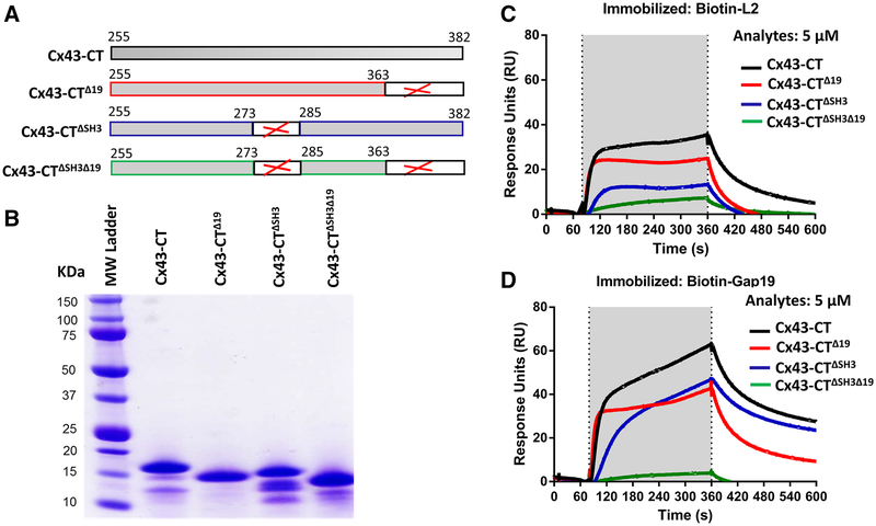
Fig. 2.
The last 19 amino acids and the SH3-binding domain are two regions within the Cx43-CT that participate in interactions with the L2 or Gap19 region. a Schematic representation of the different Cx43-CT proteins used in this study. b GelCode Blue-stained SDS-PAGE gel showing the purified Cx43-CT, Cx43-CTΔ19, Cx43-CTΔSH3 and Cx43-CTΔSH3Δ19 proteins after GST removal. 5 μg of protein was loaded per lane. c, d Sensorgrams obtained for Cx43-CT, Cx43-CTΔ19, Cx43-CTΔSH3 and Cx43-CTΔSH3Δ19 applied as an analyte at 5 μM to streptavidin-coated sensor chips loaded with biotin-L2 (c) and biotin-Gap19 (d). In both cases, the presented resonance units (RU) signals were obtained by subtracting the non-specific binding of the proteins to biotin-L2 reverse. The application of the analyte is indicated by the gray region in the sensorgram

