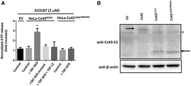
Fig. 7.
TAT-SH3 fails to restore the activity of Cx43ΔGap19M242. a HeLa cells transiently expressing EV, Cx43M242 or Cx43ΔGap19M242 were exposed to 2 μM A23187. ATP release was determined in the presence of vehicle (control), TAT-SH3, TAT-SH3 mutant and TATSH3 + TAT-L2, all provided at 100 μM. We used empty vector-transfected HeLa cells to determine the background ATP release in the absence of exogenous Cx43. Control indicates that the cells were only treated with A23187 and vehicle but not with peptide. Data show ATP release as fold induction of baseline values (n = 4). b An immunoblot stained with anti-Cx43 (E2) (upper blot) or anti-β-actin (lower blot) for lysates (30 μg of lysate/lane) obtained from HeLa cells transfected with empty vector (EV), Cx43, Cx43M242 and expression of Cx43ΔGap19M242. The left arrow indicates Cx43, while the right arrow indicates Cx43M242 and Cx43ΔGap19M242. The less than sign indicates a non-specific band also present in lysates obtained from empty vector-transfected HeLa cells (HeLa-EV)

