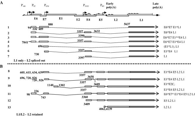
Fig. 4.
Schematic diagram of the transcripts identified by 5′RACE. (A) A diagram of the linearised virus genome. Open boxes; coding regions, arrows; promoter positions, chevrons; approximate positions of the primers used in 5′RACE. Early poly(A) and vertical bar: early polyadenylation site, late poly(A) and vertical bars: late polyadenylation sites. (B) Structures of the cDNA species identified. Grey bars: coding sequences, lines: introns spliced out. Numbers indicate the genomic positions of the 5′ ends of the cDNAs and the splice donor and acceptor sites.

