
Fig. 2.
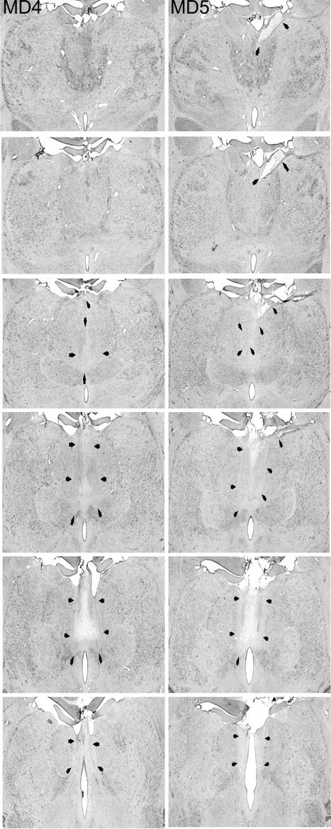
MDmc lesions cont. The two columns show photomicrographs of the MDmc lesions for MD4 and MD5 corresponding as closely as possible to the each of the schematic diagrams shown in Figure 1. Arrowheads indicate the site of each lesion.
Official websites use .gov
A
.gov website belongs to an official
government organization in the United States.
Secure .gov websites use HTTPS
A lock (
) or https:// means you've safely
connected to the .gov website. Share sensitive
information only on official, secure websites.
MDmc lesions cont. The two columns show photomicrographs of the MDmc lesions for MD4 and MD5 corresponding as closely as possible to the each of the schematic diagrams shown in Figure 1. Arrowheads indicate the site of each lesion.