
Figure 1.
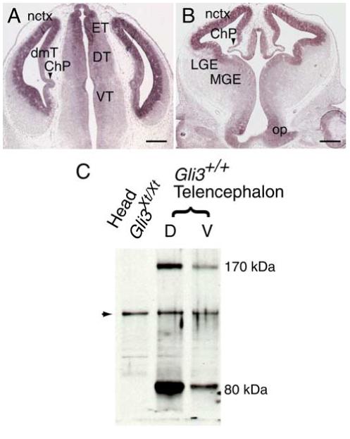
Gli3 expression in E12.5 forebrain. A, B, In situ hybridization in coronal sections of the diencephalon shows expression of Gli3 mRNA in the epithalamus (ET) and dorsal (DT) and ventral (VT) thalamus and in the optic chiasm (op). In the telencephalon, Gli3 is expressed in the neocortex (nctx), dorsomedial telencephalon (dmT), and the lateral (LGE) and medial (MGE) ganglionic eminences and is absent from the choroid plexus (ChP), indicated by an arrowhead. Scale bars, 250 μm. C, Western blot analysis of Gli3 protein in E12.5 wild-type and Gli3Xt/Xt tissue. In dorsal (D) and ventral (V) wild-type telencephalic tissue, both the long (170 kDa) and short (80 kDa) Gli3 isoforms are present, although their relative amounts differ. Both isoforms are absent in Gli3Xt/Xt whole-head protein extracts. The arrowhead indicates a nonspecific band that is used as an internal loading control.