
Figure 3.
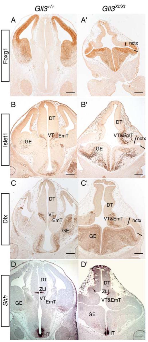
Forebrain marker analysis in wild-type (Gli3+/+) (A-D) and Gli3 mutant (Gli3Xt/Xt) (A’-D’) E12.5 coronal sections. Foxg1 protein expression delineates telencephalic tissue in wild type (A) and mutant (A’). The area between the lines corresponds to Mash1-negative neocortex (nctx) in the mutant (see Fig. 2G’, H’). Islet1 and Dlx proteins are both found in the ventral telencephalon [ganglionic eminences (GE)] in both wild type (B,C) and mutant (B’,C’). They are also observed in the ventral thalamus (VT) in wild type and within a region in the mutant that corresponds to the ventral thalamus and eminentia thalami (EmT). Note that neither Islet1 nor Dlx proteins are expressed in neocortex of the mutant (area between the lines in B’, C’). Shh mRNA is expressed in the ZLI in wild type (D) and mutant (D’), defining the border between the dorsal thalamus (DT) and ventral thalamus. Note that the orientation of the Shh-positive ZLI is perpendicular to the main axis in the wild type sbut not in the mutants. Shh mRNA expression is also observed in the hypothalamus (HT) in both wild type and mutant. Scale bars, 250 μm.