
Figure 6.
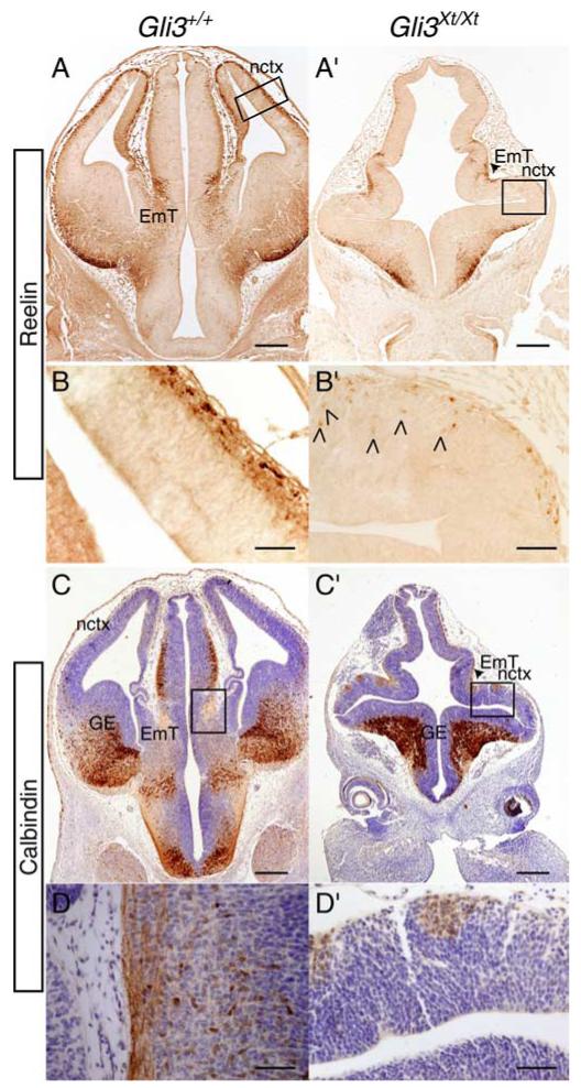
Reelin and calbindin immunoreactivity in E12.5 coronal wild-type (A-D) and Gli3Xt/Xt (A’-D’) forebrain sections. Cajal-Retzius cells in the wild-type neocortex (nctx) are labeled with reelin (A, B). The few reelin-positive cells in the mutant neocortex (A’, B’) do not correspond to the patchy areas devoid of Foxg1 staining (see Fig. 5A’). Arrowheads (B’) indicate reelin-positive cells that are not located in the most outer layer of the Gli3Xt/Xt neocortex. Reelin also labels the eminentia thalami (EmT) in both wild type (A) and mutant (arrowhead in A’). Calbindin stains cell somata and fibers in the ganglionic eminences (GE) and fiber sin EmT in both wild type (C, D) and mutant (arrowhead in C’). Adjacent to the eminentia thalami in the mutant, small clusters of calbindin-positive fibers (C’, D’) are present in the region of the neocortex. Sections in C, C’, D, and D’ are counterstained with cresyl violet. Note that D and D’ are serial sections from the same specimens as in Figure 5, A,C,A’, and C’, respectively. B,D and B’, D’ are magnifications of the boxed areas in A, C and A’, C’, respectively. Scale bars: A, A’, C, C’, 250 μm; B, B’, D, D’,50 μm.