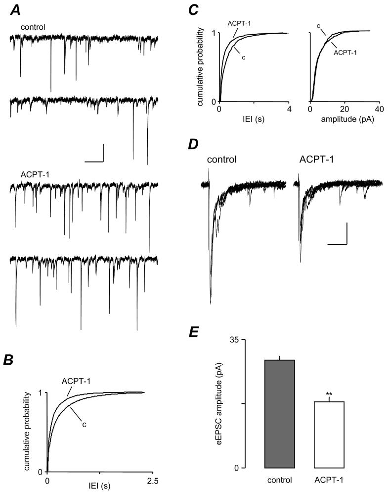
Figure 1.
ACPT-1 differentially modulates spontaneous and evoked EPSCs. A. Voltage clamp recording from a layer V neurone illustrating the increase in sEPSCs elicited by ACPT-1. B. Cumulative probability analysis of interevent interval of sEPSCs in pooled data (n=10). The shift to the left of the distribution in ACPT-1 reflects the increase in frequency compared to control (c). C. Pooled data of cumulative probability analysis of mEPSCs showing the persistence of the increase in frequency with no significant change in amplitude (n=7). D. eEPSCs (5 events superimposed) recorded in the same neurone shown in A, illustrating the concurrent reduction of AP-dependent-release. E. Summary data for eEPSC changes in the same set of neurones as in B, showing the consistent reduction in eEPSCs concurrent with the increased frequency of sEPSCs. Shows cumulative probability plots of IEI for sEPSCs for these neurones and the bar chart summarizes the effect of ACPT-1 on mean peak amplitude eEPSCs. Scale bars represent 15 pA and 500 ms in A, 30 pA and 30 ms in D.

