
Fig. 4.
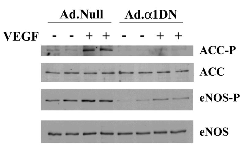
The effects of infection with Ad.α1DN on VEGF-stimulated ACC and eNOS phosphorylation.
HAECs were infected with 25 Pfu/cell of Ad.α1DN or Ad.Null 24 h prior to experimentation. Subsequently, HAEC lysates were prepared from cells incubated in the presence or absence of 10 ng/ml VEGF, resolved by SDS PAGE, transferred to nitrocellulose and probed with the antibodies indicated. Specific band intensities were quantified using NIH Image software. Representative immunoblots are shown, repeated with similar results on 4 different samples of lysates.