
Figure 1.
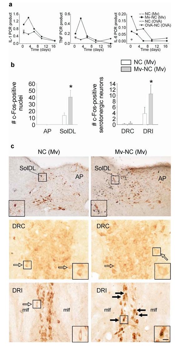
Influence of M. vaccae or its derivatives on pulmonary cytokine mRNA expression, c-Fos expression in the nTS and c-Fos expression in serotonergic neurons in the dorsal raphe nucleus. (a) Graphs illustrate mean levels, relative to β-actin, of pulmonary IL-1β (IL-1), TNFα (TNF), and IL-6 mRNA expression 12 h, and 3, 6, 10, and 17 days following i.t. injection of M. vaccae antigen in M. vaccae-preimmunized mice (•) relative to cytokine mRNA expression following i.t. injection of vehicle in M. vaccae-preimmunized mice (○) (12 h time point only), as well as, for comparison, cytokine mRNA expression at the same time points following i.t. injection of ovalbumin coupled to nitrocellulose beads in ovalbumin/alum-preimmunized mice (▼) or vehicle in ovalbumin/alum-preimmunized mice (▽) (12 h time point only). (b) Bar graphs illustrate the mean number (± SEM) of c-Fos-ir nuclei in the AP, SolDL, DRC, and DRI in Experiment 1. (c) Photographs illustrate nuclear c-Fos immunostaining (blue-black) 12 h following i.t. injection of M. vaccae in M. vaccae-preimmunized mice in the area postrema (AP) and dorsolateral part (SolDL) of the nucleus of the solitary tract (nTS) (top two photographs) and serotonergic neurons in the caudal (DRC; middle two photographs) and interfascicular parts (DRI; bottom two photographs) of the dorsal raphe nucleus (DR). Tyrosine hydroxylase immunostaining (brown) was used to aid in identification of neuroanatomical subdivisions of the nTS. Tryptophan hydroxylase immunostaining (brown) was used to identify serotonergic neurons in the DRC and DRI. (⇨) c-Fos-immunonegative serotonergic neurons, (➙) c-Fos-immunopositive serotonergic neurons. Small black boxes in (c) indicate regions shown at higher magnification in insets. Scale bar, (c) top row, 100 μm; (c) middle and bottom rows, 25 μm; (c) insets, 12.5 μm. Abbreviations: AP, area postrema; DRC, dorsal raphe nucleus, caudal part; DRI, dorsal raphe nucleus, interfascicular part; IL-1, interleukin-1β; IL-6, interleukin-6; mlf, medial longitudinal fasciculus; (Mv), preimmunization with s.c. injections of heat-killed M. vaccae; Mv-NC, i.t. challenge with sonicated heat-killed M. vaccae coupled to nitrocellulose beads (M. vaccae antigen); NC, i.t. challenge with nitrocellulose beads; SolDL, nucleus of the solitary tract, dorsolateral part; TNF, tumor necrosis factor-α. *P ≤ 0.05, compared to M. vaccae-preimmunized, vehicle-injected controls, Fisher’s Protected LSD test.