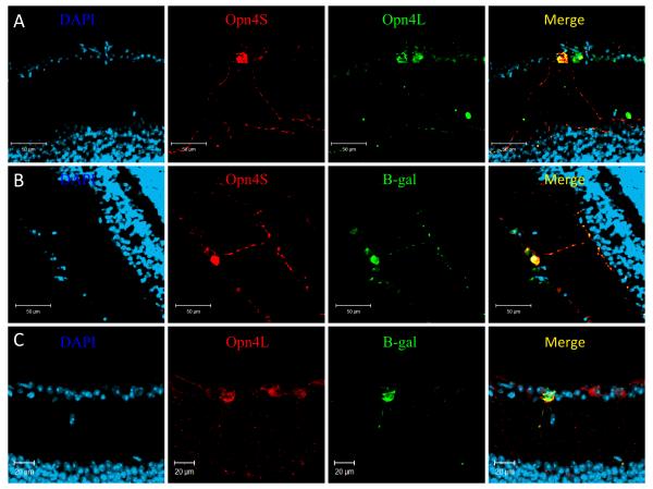
Figure 8. Immunolabelling of the mouse retina shows a differential pattern of expression for Opn4L and Opn4S.
A) Double labelling with Opn4L (green) and Opn4S (red) identified two subsets of pRGCs, those expressing both Opn4L and Opn4S and a second subset of cells expressing only Opn4L. B) Double labelling with β-gal (green) and Opn4S (red) shows a 100% overlap of expression, with all cells positive for both β-gal and Opn4S. C) Labelling with β-gal (green) and Opn4L (red) reveals a sub-set of Opn4L positive cells that lack detectable β-gal expression. For all images DAPI = blue.

