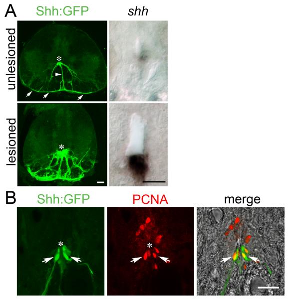
Fig. 3.
Increased expression of shha and proliferation of Tg(shha:gfp)+ ependymo-radial glial cells in the ventricular zone of the lesioned spinal cord. Cross sections are shown of whole spinal cross sections (Tg(shha:gfp) in A only) or just for the area around in the ventricle at high magnification (all other images). Asterisks indicate the position of the ventricle. A: Expression of shha mRNA is increased in the ventral-most position of the ventricle and Tg(shha:gfp)+ ventral ependymo-radial glial cells are more numerous at 2 weeks post-lesion. B: Tg(shha:gfp)+ ependymo-radial glial cells actively proliferate at 2 weeks post-lesion, as indicated by double labeling with PCNA antibodies (arrows). Bars in A = 25 μm, in B = 25 μm.

