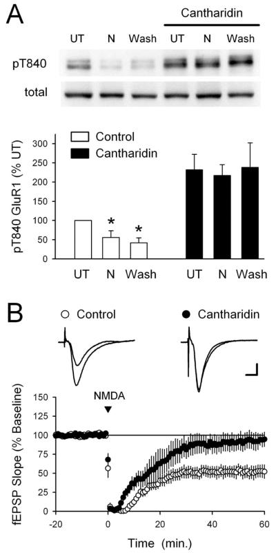
Figure 7.
Blocking PP1 and PP2A inhibits NMDA-induced dephosphorylation of GluR1 at T840. A, Slices from the same animal were maintained in high Ca 2+-ACSF either alone or in the presence of the PP1 and PP2A inhibitor cantharidin (10 μm). Under control conditions, GluR1 phosphorylation at T840 was significantly decreased both immediately after NMDA application (20 μm for 3 min; N) and 40 min after NMDA washout (Wash; *p < 0.05 compared with untreated controls; n = 5). Basal levels of T840 phosphorylated GluR1 were significantly increased in cantharidin-treated slices ( p < 0.01 compared with UT slices bathed in high-Ca 2+ ACSF alone), and both the initial and persistent NMDA-induced dephosphorylation of T840 were completely blocked. The immunoblots show phospho-T840 GluR1 and total GluR1 levels from a representative experiment. Error bars indicate SEM. B, Chem-LTD is inhibited in cantharidintreated slices. Sixty minutes after NMDA application (20 μm for 3 min), fEPSPs were depressed to 52 ± 8% of baseline in vehicle control experiments (open symbols; 0.1% DMSO; n = 6) and were 93 ± 10% of baseline in cantharidin-treated slices (filled symbols; n = 5; p = 0.01 compared with control). Slices were continuously bathed in ACSF containing 10μm cantharidin for at least 1 h before an experiment.

