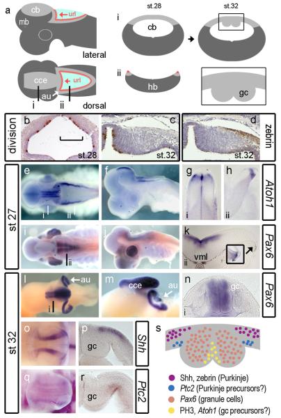
FIGURE 7. Development of granule cells in the shark cerebellum.
Schematic dorsal and lateral views (a) of the shark cerebellum show the relationship of the cce to the url whose lateral extensions comprise the cerebellar auricles (au, white arrow). Levels of transverse sections through the cerebellum (i) and hindbrain are indicated (ii) on the dorsal plan. Schematic profiles for these sections are shown for a st.28 embryo (middle). At st.32, in schematic transverse section (right), the cerebellar midline shows prominent thickenings (boxed area) comprising accumulated postmitotic granule cells (gc) (Meek, 1992b; Nieuwenhuys et al., 1998; Rodriguez-Moldes et al., 2008). Cell proliferation (brown: phosphohistone H3-positive) in the neural tube at the level of the presumptive cerebellum at st.28 (b) and unilaterally at the dorsal midline st.32 (c). The medial extent of the Zebrin-positive domain of Purkinje cells coincides with the boundary of the midline eminences (d). At st.27, Atoh1 in wholemount dorsal (e) and lateral (f) view. Transverse sections indicated in a show Atoh1 adjacent to the cerebellar midline (g) and at the rhombic lip (h). By contrast, Pax6 in dorsal (i) and lateral (j) view is restricted to the ventral hindbrain and absent from the rhombic lip (k) until st.28 (inset). By st.32, dorsal (l) and lateral (m) views show that Pax6 is expressed in the cerebellar midline nuclei and auricles. Transverse section through the cerebellar midline shows Pax6 is restricted to granule cell bundles. At the same stages, Shh (o) is expressed in lateral sagittal stripes either side of granule cells, which in transverse section (p) is restricted to the pialward cells. In wholemount, faint Ptc2 expression (q) maps to that of Shh. In section, Ptc2 is expressed in the ventricular layer (r). These expression domains are summarised (s) in a schematic diagram a mid-cerebellar transverse section of the dorsal midline.

