Fig. 1.
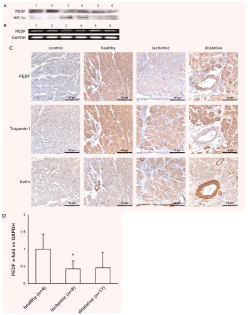
PEDF expression in human cardiac tissue. PEDF and HIF-1α determination by Western blot of homogenized cardiac tissue isolated from two healthy hearts (lanes 1 and 2) and four explanted hearts from patients suffering from ischemic (lanes 3 and 4) and dilatative (lanes 5 and 6) cardiomyopathy (A). PEDF mRNA expression in human cardiac tissue isolated from the same hearts used for Western blot in (A), e.g. two healthy hearts (lanes 1 and 2) and four explanted hearts from patients suffering from ischemic (lanes 3 and 4) and dilatative (lanes 5 and 6) cardiomyopathy; GAPDH served as a loading control (B). Immunohistochemical staining of PEDF, troponin I and actin in paraffin embedded heart tissue from a healthy heart and explanted hearts from patients suffering from ischemic and dilatative cardiomyopathy, respectively (C). mRNA was isolated from the left ventricle of healthy human hearts (n = 4) and from hearts of patients suffering from ischemic (n = 8; *P = 0.014) or dilatative cardiomyopathy (n = 17; n.s., P = 0.287); real-time PCR was performed employing specific primers for PEDF. Values represent mean values ± S.D. Values are given as x-fold of control and were normalized using GAPDH levels (D).

