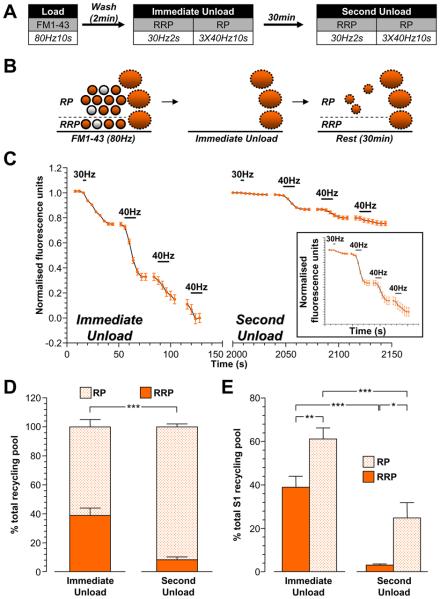
Figure 8. SVs generated from bulk endosomes specifically replenish the reserve pool.
A, Cultures were loaded with 10 μM FM1-43 using a train of 800 action potentials (80 Hz). Dye was washed away immediately after stimulation and cultures were stimulated to unload both the RRP and reserve pool (RP) after both 2 min (Immediate Unload) and a further 30 min (Second Unload). The RRP was unloaded with a train of 60 action potentials (30 Hz) and the RP was unloaded with three consecutive trains of 400 action potentials (40 Hz). B, Schematic diagram illustrating how the RRP and RP are replenished by SVs loaded with FM1-43. Solid circles represent dye-loaded SVs, clear circles represent non-loaded SVs. Solid ovals represent dye-loaded endosomes. Solid outlines represent CME-generated structures, dotted outlines represent ADBE-generated structures. C, A representative trace of the average fluorescence drop of nerve terminals is shown (Immediate Unload – S1; Second Unload – S2). Unloading stimulations are represented by bars. Traces were normalised (between 1 and 0) to the size of the S1 recycling pool (RRP + RP) for each nerve terminal. Inset displays the S2 unload normalised to the size of the S2 recycling pool. D, Proportion of the S1 and S2 RRP and RP as a percentage of their respective recycling pools is shown. E, Average proportion of the RRP and RP in both S1 and S2 expressed as a percentage of the S1 SV recycling pool (RRP - solid bars; RP – dotted bars). All experiments n = 4 ± SEM, * p<0.05, ** p<0.01, *** p<0.001, for both RRP and RP, one-way ANOVA.

