Fig. 5.

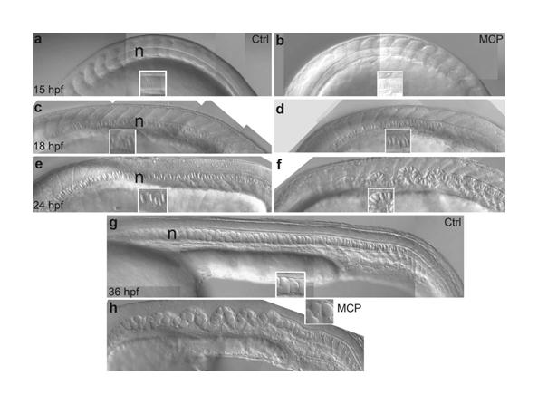
MCP 1 affects notochord morphology from late-somitogenesis stages. Lateral views of trunk of embryos treated with 200 nM MCP 1 (b,d,f,h) or left untreated (a,c,e,g) and photographed at 15 (a,b), 18 (c,d), 24 (e,f) and 36 (g,h) hpf. Insets show close-ups of notochord cells to show progressive enlargement of vacuoles. The notochord (n) appears rather normal at early stages, but rapidly becomes severely kinked during the phase of cell vacuolation (just detectable at 18 hpf (d), dramatic by 24 hpf (f)). Note in (g) and (h) the clear correlation between notochord distortion and incomplete rostrocaudal expansion.
