Figure 1.
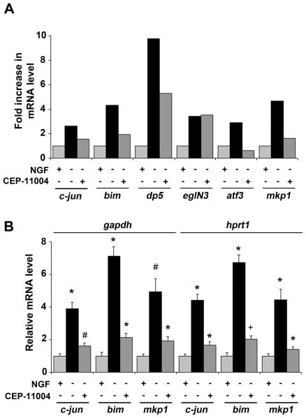
Mkp1 mRNA levels increase in sympathetic neurons after NGF withdrawal and this requires MLK activity. Sympathetic neurons from 1 day-old rats were cultured for 6 days in vitro and then rinsed and refed with medium containing NGF (+NGF), or lacking NGF (−NGF), or lacking NGF but containing 400 nM CEP-11004 (−NGF+CEP-11004) for 16 hours. RNA was then isolated using an RNeasy mini kit. A, Affymetrix Rat Exon 1.0ST microarray analysis of gene expression in sympathetic neurons at the whole gene level. c-jun, bim, dp5 and atf3 are induced after NGF withdrawal and this is reduced by CEP-11004 validating them as known targets of the MLK-JNK-c-Jun pathway. mkp1 induction after NGF withdrawal (4.51-fold) is reduced by CEP-11004 to 1.42-fold suggesting that it too may be a transcriptional target of the JNK pathway. B, c-jun, bim and mkp1 mRNA levels were measured by real time PCR and normalised to the housekeeping genes gapdh and hprt1. Similar results were obtained in both cases. The data shown represents the average of 3 independent experiments ± SEM. Statistical comparisons were made between +NGF and −NGF or −NGF and −NGF+CEP for each gene. #P <0.001; *P <0.01; +P=0.02.

