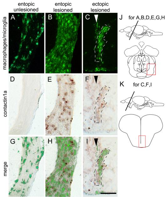
Fig. 4.
De-afferented ectopic optic tracts in the telencephalon of astray mutants display macrophage/microglial cell activation and increased contactin1a mRNA expression comparable to entopic tracts. Cross sections through the adult brain are shown as indicated in J,K. Macrophage/microglial cell immunolabeling (A-C) and contactin1a mRNA labeling (D-F) is comparable between de-afferented entopic (B, E, H) and ectopic astray optic tracts (C,F,I). Both signals are increased compared to unlesioned entopic tracts (A,D,G). Arrowheads in C,F,I indicate telencephalic midline. G, H, I shows superimposition of macrophage/ microglial cell and contactin1a mRNA labeling. Scale bar = 200 μm.

