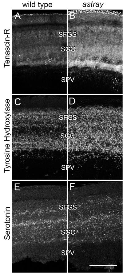
Fig. 6.
Comparison of laminar distribution of different markers in the denervated tectum at 1 week post-lesion. Cross sections through the dorsal tectum are shown (dorsal is up). Tenascin-R (A,B), Tyrosine Hydroxylase (C,D), and serotonin (E,F) immunoreactivities show comparable distribution in wild type and astray animals. However, labeling intensity of Tenascin-R and Tyrosine Hydroxylase was increased in astray mutants relative to wild type animals. For anatomical abbreviations see previous figures. Scale bar = 100 μm.

