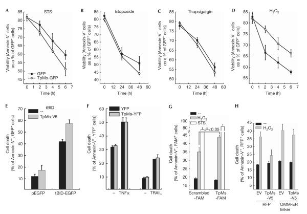
Fig 4.
Trichoplein/mitostatin protects cells from Ca2+-dependent apoptosis. (A–D) Viability of HeLa cells transfected as indicated and treated for the indicated times with staurosporine (STS, 2 μm, A), etoposide (5 μm, B), thapsigargin (1 μm, C) and H2O2 (1 mM, D). (E) Cell death of HeLa cells 48 h after transfection with the indicated and plasmids. (F) Cell death of HeLa cells transfected as indicated and treated with TNF-α (25 ng/ml plus 10 μg/ml cycloheximide) or TRAIL (50 ng/ml) for 16 h. In (A–F), data are represented as mean±s.e. (n=5). (G) Cell death of HeLa cells transfected as indicated and treated after 48 h with H2O2 or STS for 6 h. Data are represented as mean±s.e. (n=3). (H) Cell death of HeLa cells co-transfected as indicated and treated after 24 h with H2O2 for a duration of 4 h. Data are represented as mean±s.e. (n=6). EGFP, enhanced green fluorescent protein; ER, endoplasmic reticulum; EV, empty vector; FAM, carboxyfluorescein; OMM, outer mitochondrial membrane; RFP, red fluorescent protein; tBID, truncated BID; TNF, tumour necrosis factor; TpMs, trichoplein/mitostatin; TRAIL, tumour-necrosis-factor-related apoptosis-inducing ligand; YFP, yellow fluorescent protein.

