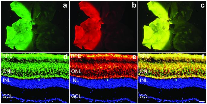
Figure 1.
Co-transduction of AAVs in wild type retinas.
Eyes of wild type mice were subretinally injected with a mixture of 1.5×10⁹ vp AAV-EGFP and 1.5×10⁹ vp AAV-DsRed. Two weeks post-injection eyes were fixed (n=5), whole mounted for imaging, then cryosectioned (12 μm) and processed for histology. Nuclei were counterstained with DAPI. a, b and c: representative whole mounts illustrating EGFP (a), DsRed (b) and overlay of EGFP and DsRed signals (c). Representative sections demonstrate significant co-expression (f) of EGFP (d) and DsRed (e) signals at the cellular level in the outer nuclear layer. In order to obtain a clearer view of the markers the DAPI (blue) signal was edited out from the ONL. RPE: retinal pigment epithelium; ONL: outer nuclear layer; INL: inner nuclear layer; GCL: ganglion cell layer. Scale bars: 1 mm (a, b and c) and 25 μm (d, e and f).

