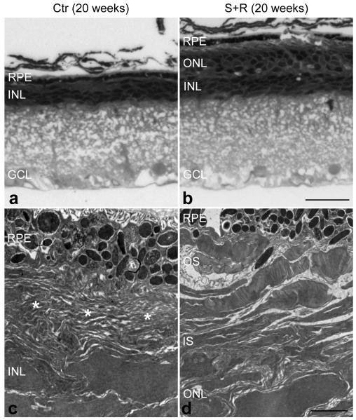
Figure 5.
Ultra-structural analysis of combined suppression and replacement treated retinas twenty weeks post-injection.
The right eyes of P5 P347S mice were subretinally injected with a mixture of 6.0×10⁸ vp AAV-S and 1.8×10¹⁰ vp AAV-R (b and d) while the left eyes were injected with 6.0×10⁸ vp AAV-C (a and c). Note that AAV-S and AAV-C co-express EGFP. Eyes were fixed, whole mounted, and transduced areas identified by EGFP fluorescence and excised. The excised retinal samples were post-fixed and processed for transmission electron microscopy (TEM). Semi- and ultra-thin sections were analysed by light microscopy (a and b) or TEM (c and d). Combined suppression and replacement therapy resulted in preservation of rod photoreceptor outer segments (OS), which extended to the retinal pigment epithelium (RPE; b and d). In contrast, in the control retina, only membranous debris (*) was detected between the RPE and the inner nuclear layer (INL) while the outer nuclear layer (ONL) was not present (a and c). IS: inner segment layer, GCL: ganglion cell layer. Scale bars: 25 μm (a and b) and 2 μm (c and d).

